Why viruses are the Bigfoot of modern medicine.
Many people have a real hard time accepting the entire ‘virus’ charade is nonsense.
This is despite modern medicine’s long, well-established track record of Making Stuff Up in order to explain the pathogenesis of diseases it doesn’t really have a clue about.
A classic case in point is the lipid hypothesis of heart disease.
By the 1950s, coronary heart disease was a leading cause of death in western countries like the US, UK and Australia. The medical establishment had no idea what the cause was. The medical establishment, of course, doesn’t like looking clueless; it enjoys an aura of all-knowingness it wishes to maintain. So it settled on the ridiculous theory that when you eat saturated fats and cholesterol, “fatty deposits” form on the walls of your arteries like mud inside a pipe. These fatty deposits get bigger and bigger, like the deadly oozing mass from the 1958 horror classic The Blob, until they block a coronary artery. This stops blood flow to your heart, which stops beating, resulting in a heart attack.
The contradictions and flaws in this simple-minded theory are so numerous, I was able to fill a book with them. I was hardly the first to point out the theory was completely untenable; criticism of the lipid hypothesis is as old as the theory itself. The anti-cholesterol juggernaut simply ignored it, or created convenient rationalizations, like labeling any evidence that contradicted the theory a “paradox”.
The lipid hypothesis was nonsense, but it spawned a massive and extremely profitable industry. Diagnostic laboratories enjoyed a steady uptick in business as doctors began ordering squillions of useless cholesterol tests. Food manufacturers cashed in wildly by marketing low-fat foods and promoting cholesterol-lowering polyunsaturate-rich plant oils. Never mind those highly refined plant oils have been shown to increase heart disease and cancer mortality - dogma is more important to people than reality, especially when there are ungodly profits to be gained from the former.
Big Pharma, of course, used the cholesterol sham to create a new class of “blockbuster” lipid-lowering drugs known as statins. Like most drugs targeting an imaginary cause, statins were ineffective and toxic, and had to be propped up by heavily manipulated research.
Needless to say, after decades of anti-cholesterol paranoia and low-fat stupidity, CHD still remains the leading cause of death for men, women, and people of most racial and ethnic groups in the US (ground zero for the war on cholesterol). In countries like the UK and Australia, coronary heart disease remains the leading killer of males, and second only to dementia as the leading cause of death in females.
Despite this dismal failure, anti-cholesterol stupidity remains deeply ingrained in these countries. When local News Corp outlets here in Australia recently reported a positive study on Coronary Artery Calcium Score tests, they described the procedure as “a CT scan of a person’s heart to flag calcium, which identifies the fatty plaques that can narrow and block arteries.” (Bold emphasis added).
For crying out loud, it’s right there in the name - the procedure tests for calcium deposits in your arteries, not fats!
Old habits - not to mention absurd beliefs - die hard.
My Partner Died, I Lost My Job, My House Was Foreclosed, My Car Was Wrecked the Week After My Insurance Expired … But What I Really Need is More Serotonin!
Then there’s the equally ridiculous “chemical imbalance” theory of depression. This stinker claims that depression arises - not from feelings of hopelessness, frustration, emptiness, social isolation and disillusionment (medicine has no cure for any of those) - but from an alleged deficiency of the neurotransmitter serotonin.
Despite having absolutely no basis in reality, the serotonin hypothesis has again made lots of undeserving people very wealthy, via the creation of an entirely new class of “blockbuster” drugs.
These drugs, known as selective serotonin reuptake inhibitors (SSRIs), inhibit binding of serotonin in the brain (and elsewhere). This supposedly leaves more 'free' serotonin available at the all-important synaptic gap between brain neurons. This increase in available serotonin, so the story goes, relieves depression and anxiety because serotonin is a "happy hormone" that makes you calm and tranquil. The basic equation behind the marketing of SSRIs is that more serotonin = less sadness and more happiness.
It is a theory that is simplistic to the point of sheer stupidity.
Despite this, Big Pharma has convinced almost the entire medical profession that depression is caused by a serotonin deficiency, and that taking SSRIs will boost serotonin levels indefinitely. Few people have stopped to consider the body may counter the increase in serotonin by decreasing its own natural serotonin production, or by decreasing the brain’s sensitivity to serotonin.
Here’s what we do know:
Depression has not decreased since the advent of SSRI antidepressants. It remains a highly prevalent condition.
Suicide rates have increased alongside antidepressant use.
The relationship between antidepressant use and suicide is a causal one, because RCT data repeatedly shows higher rates of self-harm and suicidality in the antidepressant groups.
While doing nothing to ameliorate depression beyond placebo effect in some patients, antidepressants cause a disturbing array of very real side effects. Even your sex life isn’t safe from this garbage, with SSRI drugs sometimes causing permanent loss of sexual desire and function. Gee, what a great way to make a depressed person feel better!
But not to worry, because the chemical imbalance theory is a very profitable one for Big Harma. The global antidepressant drugs market was valued at almost US $18.7 billion in 2024 and is estimated to grow at a compound annual growth rate of 7.5% from 2025 to 2034.
All based on drugs that don’t work to treat a “chemical imbalance” that doesn’t exist.
And then there’s the virus hypothesis.
Inventing a New Class of Pathogen to Explain Allegedly ‘Infectious’ Disease
The Latin word virus means "poison, poisonous liquid, sap of plants, slimy liquid, a potent juice."
A far cry from what we now call a ‘virus’.
It was in the late 1890s, with the so-called ‘discovery’ of the Tobacco Mosaic Virus, that the modern virus paradigm began to take shape.
A virus, we are now told, is a tiny, infectious pathogen containing RNA or DNA, with a protein or lipid coating.
Virologists know this, they claim, because they have repeatedly ‘isolated’ viruses.
When most people think of isolating something, they think of that something being separated from everything else.
The last thing most intelligent people assume when they hear the word ‘isolation’ is adding a bunch of other stuff to the thing that is to be isolated.
But that’s exactly what happens during the sham that is ‘virus isolation’.
When Isolation Becomes Rampant Confounding
Most viruses are so tiny, so the story goes, they can only be directly “visualized” by a transmission electron microscope. One of those will set you back at least US $500,000.
Not only will you need a very expensive and powerful microscope, but you’ll need to engage in a procedure known as “cell culturing”. This is where you take a patient sample (e.g., a swab of saliva, lung fluid, blood, poo, etc), and then mix it with a bunch of other stuff including culture medium and bovine fetal serum (which, not inconsequentially, contains RNA and probably DNA).
This is about as far from “isolation” as Australia is from Iceland, but the idiocy doesn’t stop there.
To this mix, virologists further add a cell line (which means more extraneous RNA and DNA). The cell line is deliberately chosen because of its propensity to degrade in vitro. This allows them to claim a “cytopathic effect” (cell-destroying effect) allegedly caused by whatever virus they portend to be looking for. This cell degradation is taken to be proof that a virus is in the mix, even though viruses supposedly need healthy, functioning cells to survive and replicate.
At this point, you may be asking, “where do these clowns get off on adding a bunch of other goo to a patient sample, and calling it ‘isolation’?”
Great question.
One to which virologists have a highly convenient answer. Viruses, they claim, can only replicate inside of cells. That’s why you put the patient sample supposedly containing a ‘virus’ into the mixture containing cells.
The implication here is that, to be able to visually observe a virus, it must be replicating. Which, when you stop to think about it (which most people, including virologists, clearly don’t) is a piss-poor justification for the whole cell culture charade.
Imagine saying to someone that, to observe dogs or cats or ringtail possums, you first have to put them in an environment where they can start boinking each other and bearing offspring.
They’d assume you’re a bloody weirdo.
But more on the viruses-need-to-replicate-before-you-can-see-them charade in a moment. First, we must finish our quick primer on the cell culture circus.
Virology’s standby and absolute fave cell line of choice for these virus culture experiments are Vero cell lines, which are obtained from the kidneys of African green monkeys.
A perfectly logical choice when studying things like influenza ‘viruses’ that supposedly infect human respiratory tracts, right?
While not as ubiquitous as monkey kidney cells, HeLa is another cell line popular among those perpetuating the cytopathic effect charade. This cell line is named after African-American woman Henrietta Lacks, who passed away from cervical cancer in 1951 at only 31 years of age. A lab researcher noticed that a sample of her cancer cells were unlike any others he’d ever seen: Where other cells would die, her cells doubled every 20 to 24 hours. While clearly unrepresentative of typical human cells, the durability of the HeLa line made it a favorite with researchers.
But the absurdities don’t end with using cell lines that are clearly unrepresentative of typical human cells.
Heck no. Virologists are just getting warmed up.
They also add antibiotics to the cell culture mixture, supposedly to prevent contamination. But guess what? Antibiotics are toxic to kidney cells, and constitute one of the most common causes of drug-induced nephrotoxicity.
Given that monkey kidney cells are the most popular choice in cell culture ‘isolation’ experiments, this is definitely a problem.
Just not to virologists, apparently.
Wheel Out the Electron Microscope
After ‘proving’ there is a virus by observing the so-called cytopathic effect, researchers then “visualize” the virus by further examining the mix under an electron microscope.
They look for round things with dots inside, scream “Eureka!”, and declare they have visually documented whatever virus they are supposedly ‘isolating’.
Never mind that, inside and outside of pathogen-free cells, there are lots of other round things with dots inside them known as cellular vesicles, examples of which are endosomes and exosomes.
Take a look at the pictures below.
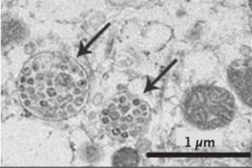
This first one is from the the New England Journal of Medicine paper of January 24, 2020, in which Chinese CDC researchers announced to the world they had isolated Sars-Cov-2.
In reality, they did nothing of the sort, as I explain in detail here.
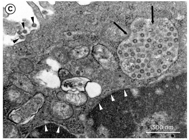
Here’s an image from the NEJM paper of cells allegedly infected with Sars-Cov-2 virions. The images show round, enclosed bodies with multiple round dots inside; those dots are allegedly Sars-Cov-2:
Now, here's an image from the June 1, 2020 Medical Journal of Australia paper in which researchers from Melbourne’s dubious Doherty Institute claimed to have performed the world’s first ‘isolation’ of Sars-Cov-2’ outside of China. Like the image above, this one also purports to show Sars-Cov-2-infected cells:
Here's an image from Korean researchers, again purporting to show cells infected with Sars-Cov-2:
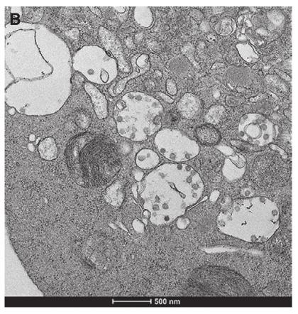
Now, feast your eyes upon the image below.
It was published by Sahoo et al in 2011, eight years before the Great COVID Con kicked off.
It is an image of exosomes - not virions - inside multivesicular bodies emanating from human hematopoietic stem cells (found in bone marrow).
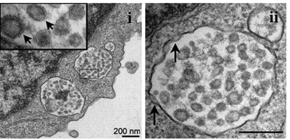
The Chinese/Korean ‘Sars-Cov-2’ virions and Sahoo et al exosomes look very similar, don't they?
What researchers are claiming to be virions are in all likelihood cellular vesicles like exosomes.
So what are exosomes? They are a type of extracellular vesicle, ubiquitous to cells.
"Extracellular vesicles (EVs) are lipid bilayer-enclosed entities containing proteins and nucleic acids that mediate intercellular communication, in both physiological and pathological conditions. EVs resemble enveloped viruses in both structural and functional aspects. In full analogy with viral biogenesis, some of these vesicles are generated inside cells and, once released into the extracellular milieu, are called ‘exosomes’. Others bud from the plasma membrane and are generally referred to as ‘microvesicles’."
"Nowadays, it is an almost impossible mission to separate EVs and viruses by means of canonical vesicle isolation methods, such as differential ultracentrifugation, because they are frequently co-pelleted due to their similar dimension. To overcome this problem, different studies have proposed the separation of EVs from virus particles by exploiting their different migration velocity in a density gradient or using the presence of specific markers that distinguish viruses from EVs. However, to date, a reliable method that can actually guarantee a complete separation does not exist." (Bold emphasis added)
-Giannesi et al, Department of Science, Roma Tre University, Italy, 2020.
This is a critical passage, because virology has long operated on the unfounded premise that centrifugation (spinning mixtures around at crazy high speeds) and exposing mixtures to density gradients (and also using filters) somehow ‘purifies’ a mixture and helps ‘isolate’ a virus. But that would only be true if there were no other entities in the mix of a similar size, shape and/or density. Needless to say, that’s a huge and unfounded leap of faith. Furthermore, you only have to look at the first two images above, both featuring centrifuged mixtures, to see there are a multitude of entities not just of similar size and shape, but of different size and shape.
The bottom line is that virologists can’t even begin to guarantee the difference between an extracellular vesicle and a virus. But not to worry - they just go ahead and claim the former is a virus any old how.
The Genome Sequencing Scam
The hopelessly confounded wankery doesn’t end there. To round out this charade, and give it the aura of diagnostic usefulness, researchers then set about determining the ‘genome’ of this ‘virus’.
Not by isolating the virus, disassembling it, and recording all its constituent nucleotides. They cannot do this, because it’s impossible to disassemble something that doesn’t exist.
So what they do instead is allegedly extract some RNA or DNA from the mixture. From this they obtain short, partial sequences of nucleotides.
They then feed these partial sequences into a computer program designed to churn out complete genomes. The computer program does as it is designed to do, and churns out what is known as an in silico genome (basically, a genome that exists on the computer).
PCR testing, a key fraud that was used to prop up the COVID scam, also works by using incomplete nucleotide sequences.
It seems that certain partial nucleotide sequences can be detected in people exhibiting similar symptoms. These people then register ‘positive’ on a test for Sars-Cov-2, or HIV, or measles, or whatever other fairy tale pathogen is being tested for.
This leads stupid people who think they are smart yet have just been destroyed in a debate with Andrew Kauffman despite resorting to every disingenuous argumentation method known to mankind (hi Steve Kirsch!) to post face-saving articles titled “How can millions of people, all exhibiting signs of COVID have whole genome sequences that match the SARS-CoV-2 reference genome if viruses don't exist?”
People like Kirsch think that because you test ‘positive’ for a virus, that in itself is proof of the virus.
First of all, just because two genomes match, it doesn’t mean we have a ‘virus’ on our hands. We just have a couple of printouts created by a computer featuring the same nucleotide codes. Virologists say it’s a virus; I say it’s the blueprint for a microscopic Ferrari. We both have the same amount of valid scientific proof for our respective contentions, which is stugatz.
As for how those two computer printouts of a ‘genome’ came to match each other - that’s easy. If you observe that particular nucleotide sequences are commonly found in people exhibiting a similar set of symptoms, then you’re all set to declare a new ‘virus’, a ‘genome’ for that ‘virus’, and an accompanying test.
This is exactly what happened in the case of Sars-Cov-2. The whole ruse was planned well in advance.
The Chinese concocted an in silico genome during their farcical ‘isolation’ escapade. The dubious Corman-Drosten team then retrieved this genome from the Internet, and custom-built a PCR test. At no time did the Corman-Drosten team utilize an actual, real live ‘virus’ to design or confirm their test.
Because the test was a sham designed to create global hysteria, Corman et al cut corners on an already flawed technology. A collaboration of twenty-two researchers (including Dr Michael Yeadon) found so many critical flaws in the test design that they called for the Corman et al article to be retracted.
Among the serious flaws identified by Borger et al:
Unusually high concentrations of primers were used.
Six unspecified positions were identified in the primers and probes, introducing “enormous variability” and rendering “the test unsuitable as a specific diagnostic tool to identify the SARS-CoV-2 virus.”
The test could not discriminate between the whole virus and viral fragments.
Corman et al provided no specific cycle threshold (Ct) value at which a sample is considered positive or negative. This left the test open to further abuse by globalist-compliant public health authorities, who were able to simply keep repeating the alternate heating and cooling cycles inherent in the PCR procedure until the test returned a positive result. This is exactly what happened during the scamdemic, where testing routinely involved absurd Ct counts in the 40s. Even Fauci admitted in a rare moment of candor, “…If you get a cycle threshold of 35 or more, the chances of it being replication-confident [i.e. accurate] are miniscule … you almost never can culture virus from a 37 threshold cycle, so I think if someone does come in with 37, 38, even 36, you gotta say, you know, it's just dead nucleotides, period.”
In line with virology’s allergy-like aversion to the use of control procedures, Corman et al employed neither a unique positive control to evaluate the test’s specificity for SARS-CoV-2 nor a negative control to exclude the presence of other ‘coronaviruses’.
As noted, the sequences on which their PCR method was based used in silico sequences derived from the Chinese. At the time of development of the test, no control material of infectious (“live”) or inactivated Sars-Cov-2 was available to the authors. There still isn’t any bonafide Sars-Cov-2, of course, but at the time they couldn’t even make a pretense of having such material on hand!Size matters, especially when it comes to entities that can only be seen with an electron microscope. I mean, it’s not like you can pull out a tape measure and declare, “yep, that’s definitely too small to be any other coronavirus!” Therefore, validation of the size of whatever the test is claiming to detect is crucial. Yet Corman et al did not perform biomolecular validation of the amplified PCR products resulting from their test. “The fact that these PCR products have not been validated at molecular level,” wrote Borger et al, “is another striking error of the protocol, making any test based upon it useless as a specific diagnostic tool to identify the SARS-CoV-2 virus.”
A subsequent paper by the same research group screened twenty papers analyzing performance of the Corman-Drosten test in “wet labs” (labs that handle biological material, instead of dry matter or pixels on a screen). Of those, 17 found problems with incorrect primer design.
The journal that published the Corman-Drosten paper, Eurosurveillance, refused to retract the article. When pressed about the paper’s incredibly suspicious peer-review and publishing timeline, Eurosurveillance flatly refused to share the peer-review notes. The journal ‘investigated’ itself and the authors - two of whom (Christian Drosten and Chantal Reusken) were also members of the editorial board of Eurosurveillance - and declared no evidence of impropriety.
Sure thing.
So when a guy like Steve Kirsch asks “How can millions of people, all exhibiting signs of COVID have whole genome sequences that match the SARS-CoV-2 reference genome if viruses don't exist?”, he’s asking the wrong question.
The real question is, “How can millions of people, all exhibiting zero signs of COVID and falling squarely into the asymptomatic category, along with papaya, quail and goat, have whole genome sequences that match the Sars-Cov-2 reference genome when Sars-Cov-2 doesn't even exist?”
The answer, of course, is that the tests for Sars-Cov-2 are highly flawed and non-specific garbage.
Wanted Alive or Dead
Okay, a quick recap. Most of what researchers claim to be viruses have only ever been seen after the bodily fluids or excretions they have allegedly infected were first mixed into a ‘cell culture’ - one typically containing culture medium, bovine fetal serum, African green monkey kidney cells, and antibiotics.
When we ask why the need for this cell culture caper, virology replies that viruses can only replicate inside of cells.
The official tale gets a bit murky and contradictory here - as elaborate lies often do. Some researchers claim viruses cannot replicate outside of cells but they can still survive, even on inert surfaces, at least for a while.
Researchers from the CDC and Fauci’s NIAID claimed in a leading medical tabloid that the ‘Sars-Cov-2 virus’ can survive on plastic and stainless steel “up to 72 hours after application.”
Which would mean your super-duper microscope that costs more than two Lamborghini Huracans shouldn’t have any problem taking a happy snap of a living Sars-Cov-2 virion outside of cell culture.
But no-one has ever done that.
Hmmm.
Others researchers claim that, not only do viruses fail to replicate outside of cells, but they cannot function at all outside of cells.
Well, if you’re not functioning at all, then you must be dead.
And that is exactly what many virologists, bless their eternally imaginative hearts, now claim.
Yep, they maintain that a virus is dead outside of cells, but comes alive inside of cells!
It then dies again after being “shed”, a process in which it allegedly leaves its current host, floats through the air for up to precisely 1.49 meters or 5’11” courtesy of a cough or sneeze or even just a regular breath, before settling on a counter top, where it lies in wait for its next victim.
According to this most fanciful theory, even when a virus is lying dead, nothing but a piece of inanimate lifeless debris, it still remains highly infectious.
I smell sheer absurdity.
Because in addition to the Goodfellas Theory of Viral Replication, which holds that a virus first needs to get inside a cell and “hijack” its “cellular machinery”, we now have the Jesus Died and then Rose From the Dead Theory of Viral Spread, which holds that a virus can alternate between living and non-living!

But Houston, tenemos un problema. Adding yet another ridiculous explanation on top of the preexisting ridiculous explanations still doesn’t explain away virology’s failure to isolate a virus.
Viruses, you see, don’t vaporize in their dead state. They are still a physical entity. So why can’t researchers simply take a photo of dead virions on inert surfaces? Surely, after decades of ‘virus ‘research, they can tell the difference between dead virions and dust particles, fungi, bacteria, tiny Red Bull droplets, etc, etc?
If I see a dead snake, I still know it’s a snake - not a lizard or a piece of bungy cord. If I see a dead cockroach, I know it’s a cockroach and not a spider or a beetle.
Yet virologists, in 2025, having allegedly determined long ago what living viruses look like, still can’t tell the difference between a dead virion and a dust mite, even with their uber-powerful electron microscopes?
Or is the real reason for their inability to capture images of virions on inert surfaces due to the fact the entire virus charade is a load of codswallop?
Is it because what they are really showing us in all those electron microscope images of cell culture are not virions, but things like exosomes? Is it because they can pull this switcheroo trick in cell culture experiments, but not on plastic or stainless steel surfaces where there are no cellular organelles to sneakily pass off as ‘virions’?
The virus paradigm is a fabrication by the world of science to explain things that scientists can’t otherwise explain. Just like the lipid hypothesis of heart disease and the serotonin/‘chemical imbalance’ theory of depression.
The virus theory came about to explain diseases of a seemingly “infectious”, “contagious” nature, for which they couldn’t identify a bacterial culprit.
And, just like the lipid hypothesis and chemical imbalance’ theory, the virus charade continues to provide a convenient excuse for the ongoing global administration of highly profitable and highly toxic drugs.
But how did this whole virus fairy tale gain traction in the first place?
Stay tuned, for this will be discussed in the next installment.
Until then, here’s some recommended reading material:


Leave a Reply