The absurd laboratory procedure that supposedly proves the presence of a virus, but does no such thing.
To ‘prove’ the existence of viruses, the dark art of virology often relies on something known as the “cytopathic effect”. This occurs during the farce known as ‘virus isolation’, in which no virus is ever isolated. Instead, a sample of bodily fluid or excrement is taken from a patient, and mixed with a host of other items including (typically) monkey kidney cells, culture medium, bovine fetal serum and antibiotics.
The antibiotics are thrown in supposedly to kill bacteria, but as you’re about to learn, serve a far more useful purpose for virologists.
This multifaceted mix is known as a “cell culture”.
To anyone with a functioning brain, it should be obvious this is not isolation. Mucus itself contains numerous components including mucins, glycoproteins, proteoglycans, lipids, proteins, and DNA. It can also contain pathogenic bacteria, fungi and trapped foreign matter such as dust, pollution particulates and allergens.
This is simply thrown into the cell culture mix, which further contains a bunch of other ingredients.
So in order to ‘isolate’ a virus, virology uses a process that is the exact opposite of isolation. It’s classic Orwellian playbook methodology, in which words are used in a manner that totally contradicts their true meaning, and everyone just nods along like nothing untoward is taking place.
The cytopathic effect charade kicks off when researchers notice the cells in the culture have begun to deform and die. This cytopathy is taken as proof there is a virus in the mixture.
Let the Cherry-Picking Begin
It must be noted there are a very wide range of cell lines researchers could use during these experiments. In experiments ‘isolating’ influenza viruses like the mythical Sars-Cov-2, the natural choice would be human respiratory cells because, according to Uncle Sam’s NIH, Sars-Cov-2 “infects the cells along the airways. It invades the cells in part by attaching to a specific cell receptor, called ACE-2, found on each cell. The virus uses ACE-2 as a doorway into the cell.”
If you know anything of value about the human body, the ACE-2 receptor story tells you right off the bat the official Sars-Cov-2 story is a complete and utter crock.
Cell receptors are found either on the cell membrane, or inside the cell. The official Sars-Cov-2 fairy tale claims that the virus engages with ACE-2 receptors on the cell membrane.
Now, the thing to remember about cell receptors is they are very specific. They’re picky little bastards, and they have to be, because they are part of the mechanism that ensures the wrong things don’t enter our cells.
To quote ScienceFacts.net: “Like every lock has a specific key, every receptor molecule only recognizes and responds to a particular signaling molecule (ligand).” (Bold emphasis added)
Virions, meanwhile, allegedly contain RNA or DNA surrounded by a protein or lipid coating. Virions need access to host cells to survive, so the story goes, which means they die outside of the body, although NIH researchers claim Sars-Cov-2 can survive on inert surfaces for up to 72 hours. Which makes you wonder why, instead of enacting the cell culture voodoo, no-one has simply taken an electron micrograph of these isolated virions on an inert surface, to which the answer is Sars-Cov-2 doesn’t exist.
But I digress.
Meanwhile, angiotensin-converting enzyme 2 (ACE-2) plays an important role in lowering blood pressure. It has a specific receptor on the cell membrane, known as membrane bound angiotensin-converting enzyme 2 (mACE2).
So the $64,000 question is why on Earth would a receptor that has a sole purpose - binding with ACE-2 and allowing it into cells - bind to some bizarre molecule of RNA coated with proteins that alternates between dead and alive?
Imagine you are a doorman for a club that, unlike other dive bars in the district, has established a friendly, relaxed vibe with well-dressed and well-behaved patrons. As doorman, your job is to make sure things stay that way. You have very specific instructions from the owner as to what type of patrons are allowed into the club, and these instructions have proved instrumental in keeping away trouble-makers.
One night, a couple of strange-looking zombies walk towards the door. They barely look human and look like they just came back to life after lying dead for several days in a NIH lab.
“Hello, hello,” you mutter to yourself, “what do we have here?”
As they approach, intuition honed from years of working the doors tells you these characters are up to no good.
You block the entrance to the club and politely tell the zombies, “sorry gentlemen, but we’re full.”
One of them ignores you and tries to push past. You grab him, and ‘guide’ him away from the door.
“The venue is full,” you reiterate, this time in a notably firmer tone of voice.
The other zombie rushes towards you, only to be abruptly stopped in his tracks as your right Crockett & Jones sinks into his groin.
As he writhes in agony, the other zombie glares at you, contemplating his next move.
“Fellas, we can do this all night, but you ain’t coming in. Now why don’t you lads go home, have a wash and a warm tea, and get an early one?”
The zombies finally take a hint and slink away.
What virology wants you to believe is that Sars-Cov-2, which has never been isolated, rolls up to the cell door made for ACE-2. Despite looking nothing like ACE-2, mACE-2 looks this strange, totally unfamiliar molecule up and down and then immediately says, “yeah, no worries mate, in you come! Make yourself at home!”
Vaffanculo.
Now hold that ACE-2 thought for a moment, while we discuss the next fatal flaw in the cell culture wank. Alleged influenza viruses like Sars-Cov-2 allegedly infect human beings by attaching to ACE-2 receptors in their lungs and airways.
So the natural choice of cell line for these cell culture experiments would of course be those taken from the lungs or respiratory tracts of human beings.
Yet virologists never use lung or respiratory tract cells when studying respiratory ‘viruses’.
Instead, they almost always use monkey kidney cells.
Not only that, they typically use a specific type of monkey kidney cell line, known as Vero, taken from African green monkeys.
To listen to virologists, the reason they use Vero cells when isolating Sars-Cov-2 is because these cells have high levels of ACE-2 receptors, which makes them ideal for attracting the virus.
That’s complete bollocks. Here is the real reason virology has such an attachment to Vero cells:
Vero cells give virologists the results they are looking for. That’s because Vero cells are especially prone to the cytopathic effect, so they help ensure that an ‘isolation’ experiment will be a success and ‘prove’ the existence of a virus.
Virologists never tell you that antibiotics are well-known to be toxic to kidney cells.
Little wonder they can produce the cytopathic effect upon demand.
Rigging Experiments to Produce the Cytopathic Effect
In March 2020, ‘scientists’ from the criminal enterprise known as the US CDC claimed they had 'isolated' Sars-Cov-2.
Using Vero cells of course.
They tried "to infect and replicate" the Woohoo virus in several common primate and human cell lines, including human adenocarcinoma lung cells (A549), human liver cells (HUH7.0), and human embryonic kidney cells (HEK-293T), in addition to Vero E6 and Vero CCL81 cells, and even "an available big brown bat kidney cell line (EFK3B)."
What happened when they tried to ‘isolate’ Sars-Cov-2 in these other cell lines?
Stugatz.
"No cytopathic effect was observed in any of the cell lines except in Vero cells ... In contrast, both HUH7.0 and 293T cells showed only modest viral replication and (human lung) A549 cells were incompatible with SARS-CoV-2 infection."
Stop and read that last quote again carefully.
They admit - not publicly of course, but deep in a journal paper that few people will carefully read - that they could not get the mythical Sars-Cov-2 to infect human lung cells.
This super dooper deadly virus, that was allegedly so dangerous authorities had to put the entire planet under house arrest and bash people for not wearing masks, could not be made to infect lung cells from human beings. You know, the very cells in the very organs of the very species that was supposedly at such dire risk of catching the Woohoo virus.
Virologists don’t prefer Vero cells because they have a high concentration of ACE-2 receptors or any other such nonsense. They use Vero cells because these cells give them the results they are after.
That’s why they use Vero cells when isolating all manner of ‘viruses’, not just Sars-Cov-2.
Let’s take the example of measles, and let’s go all the way back to 1954 when Enders and Peebles published the seminal measles ‘isolation’ study that really got the cytopathic effect charade rolling.
The authors discussed how they tried to get the cytopathic effect ruse going with the following cell lines: Human embryonic lung, human embryonic intestine, human embryonic skin and muscle, human foreskin, human uterus, human kidney, embryonic chick tissue, rhesus monkey kidney and rhesus monkey testis.
In other words, to create the cytopathic effect that supposedly demonstrates isolation of a ‘virus,’ they tried a vast array of cell lines - everything from penis trimmings to monkeys’ balls.
Yet they settled on just two cell lines: Human kidney and monkey kidney cells.
Why?
Because only kidney cells would allow the results they were after. Enders and Peebles used both penicillin and streptomycin in their cell culture experiments, and kidney cells were the only cell lines that could be relied upon to deform and die in response to their procedures.
Virology is a fraud, one that has made an accepted scientific practice out of blatantly moving the goal posts in order to attain the desired results.
But Wait, It Gets Dumber
After observing the cytopathic effect in their cherry-picked monkey kidney cells, researchers yell “bingo!”
They have allegedly just ‘isolated’ a virus. And nothing keeps those government grants coming in like discovering a new virus!
Not to mention nothing justifies rolling out a new class of toxic and highly profitable ‘vaccines’ like a new virus!
But as a 5 year old could probably tell you, the researchers haven’t isolated diddly squat. They have observed cells dying, but that’s not the same as observing a virus.
So the next step is to put the mix allegedly showing virus-induced cytopathy under an electron microscope in order to visually observe the virus.
Most people visualize a cell as a round or oval thing with a membrane, some fluid inside, and a nucleus floating around in that fluid. If you’re into exercise, you might also picture a few mitochondria floating around inside the cell as well.
The truth is that a cell is a complex structure, one busier than a Myers store during Boxing Day sales. In addition to the membrane, nucleus and mitochondria, you’ve got a cytoskeleton and all sorts of structures of different shapes known as organelles.
You’ve got ribosomes, smooth and rough endoplasmic reticulum and Golgi Apparatus, which together synthesize, modify, package and sort proteins and lipids. So in addition to organelles, you have a wide array of proteins and lipids inside the cell.
You’ve got lysosomes, which break down waste materials and cellular debris. You’ve got peroxisomes, involved in metabolism and detoxification.
You’ve got microtubules, which look like, well, thin tubes. Kinda like the Mosaic Tobacco Virus.
Then you’ve got vesicles, which are found not only inside cells, but outside them as well. Surrounded by a lipid bilayer and filled with cytoplasm, vesicles have a range of functions including transporting stuff from outside the cell to the inside (endocytosis) and vice-versa (exocytosis).
In images captured by electron microscopy, endosomes and exosomes often appear as round structures with smaller round structures inside them.
Here are exosomes from human CD34+ stem cells. For reasons that will be obvious shortly, you should be aware these images were published in 2011 - eight years before the COVID-19 con kicked off.
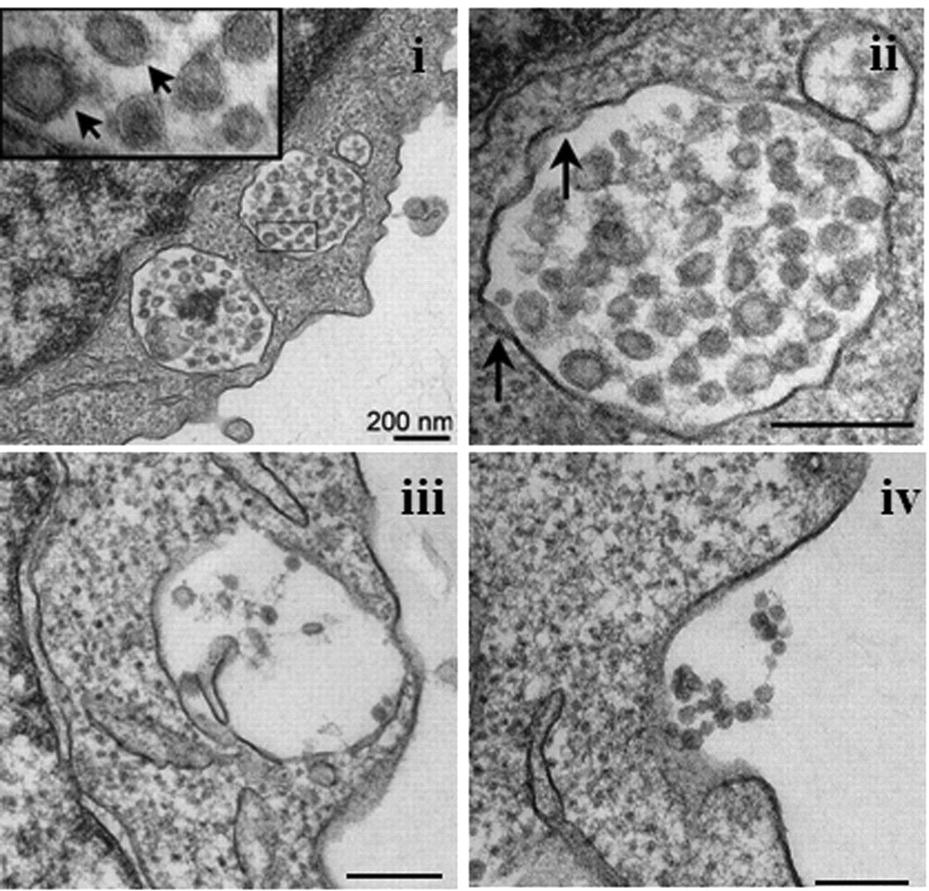
Below are images of endosomes.
Now, take a look at these images of what researchers claim to be Sars-Cov-2 virions.
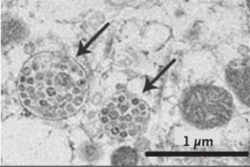
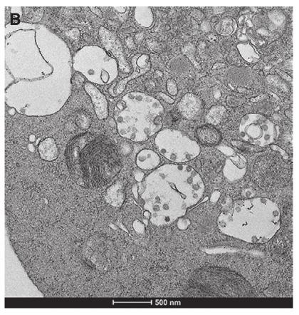
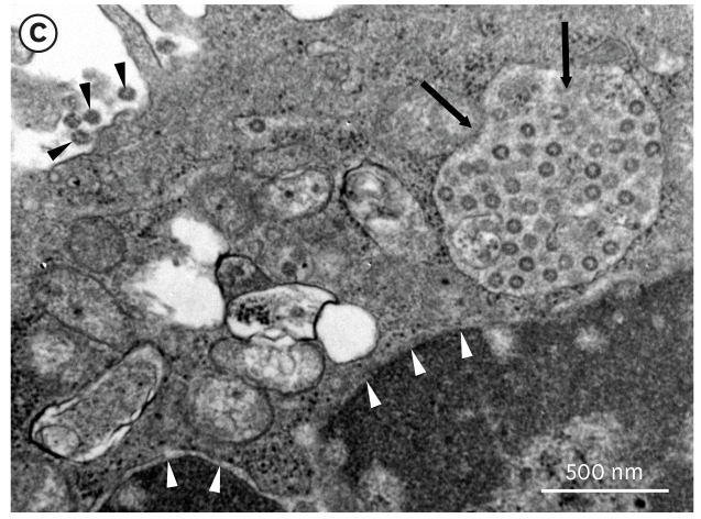
They look a lot alike, don’t they?
There are a myriad of structures and substances inside and outside a cell, that, if you were so motivated, you could claim to be ‘viruses’. In the case of Sars-Cov-2, what researchers claim are virions in fact look just like intracellular and extracellular vesicles.
Virology’s Lack of Control
So here we are, with virology claiming the specific cell receptor for ACE-2 is in fact a cellular floozy who hooks up with dodgey-looking strangers, and that what look just like cellular vesicles are in fact Sars-Cov-2 virions.
One of the prerequisites for giving this clown show any credence would be valid control procedures, which are considered par for the course in other areas of science.
Control procedures involve holding all other variables constant, in order to make sure what is being perceived as the presence of a virus is not something else. Because the cytopathic effect is heralded as ‘proof’ of a virus in cell culture, control procedures would involve performing the cell culture procedure with:
cultures with no samples
cultures with samples from healthy patients
cultures with samples from the ‘sick’ patients who yielded the allegedly ‘positive’ samples after they are fully recovered and symptom-free.
Ideally, all three procedures would be performed. In reality, viral ‘isolation’ experiments sometimes feature control procedure 1, but almost never 2 or 3.
To add to the absurdities, these isolation studies typically involve as little as one supposedly infected patient. The Chinese experiment that supposedly first isolated Sars-Cov-2 was performed using a mere three samples. The Doherty researchers who claim to be the first to have isolated the Woohoo virus outside of China used a grand total of one (1) patient - a guy who conveniently flew into Melbourne from Wuhan and suffered what was probably bacterial pneumonia (it responded to antibiotics and low-flow oxygen treatment).
Virology doesn’t like implementing valid control procedures, and a recent preprint by Matthew North, of Matthew’s Substack, goes a long way towards explaining why. Matthew’s paper is titled “The Non-Specificity of Cytopathic Effects: Implications for Virological Research and Public Health” and can be downloaded here.
He lists thirty-one studies that show the cytopathic effect to be a fatally-flawed paradigm. I won’t go through them all (read the paper and follow the links), but will discuss a few here to illustrate why the cytopathic effect charade is a load of horse manure.
Numerous of the discussed studies featured control cultures with no patient sample.
First on the list is the seminal 1954 paper by Enders and Peebles, in which the duo announced the alleged isolation of the measles virus.
Enders and Peebles performed the cell culture voodoo, and of course claimed to have observed cytopathic effect. Which, they further declared, was proof of the measles virus.
But there was a problem.
They implemented a control procedure by creating “an uninoculated culture of monkey kidney cells” that contained no patient sample.
And guess what?
It too demonstrated the cytopathic effect!
Critically, they wrote: “The cytopathic changes it induced in the unstained preparations could not be distinguished with confidence from the viruses isolated from measles.”
That’s a big deal, one that again threatened to undermine their entire isolation charade, but they quickly built an escape hatch.
They brushed off the finding by claiming they had just isolated an unidentified “agent” and that“when the cells from infected cultures were fixed and stained, their effect could be easily distinguished since the inter-nuclear changes typical of the measles agents were not observed.”
So what? That could be explained, not by a virus, but by an unidentified “agent” - not in the uninoculated preparation, but in the supposedly measles-containing culture. The uninoculated sample contained no patient sample, which automatically means the absence of a host of other substances and hence other potential triggers of “inter-nuclear changes”.
Rustigian et al 1955 reported that, “Following inoculation of the mouse-adapted strain of Hawaii dengue virus (137th mouse passage) into monkey kidney cultures, no cytopathic effects were observed for 16 days.”
Not to be deterred from getting their much-needed cytopathic effect, “fluid harvested on 12th day from 3 cultures was pooled and passed into fresh 9-day-old monkey kidney cultures.”
“With this second passage cellular changes, as described, occurred in all 5 inoculated cultures in 8 to 20 days.”
But even after finally forcing “cellular changes”, the researchers had another hiccup.
“But of 3 uninoculated control cultures held for 33 days, one revealed identical degeneration in 19 days.” (Bold emphasis added)
Whether that sentence is some kind of Masonic signal I can’t say, but they are admitting that an uninoculated culture exhibited identical cytopathy as those supposedly containing a Dengue virus strain.
So did they carefully examine this uninoculated culture to see how and why it also produced the cytopathic effect?
Nope.
“This culture, unfortunately, was discarded.”
Of course it was. When evidence doesn’t support a cherished thesis, it is often quietly relegated to the round file.

Cohen et al 1955 took samples from seven measles patients. “Transmissible agents, presumably viruses, were isolated” from two patients. No cytopathic effect was observed in samples from the other five.
“Enders and Peebles and Rustigian et al,” noted the researchers, “encountered latent virus-like agents that induce marked vacuolization and syncytial masses in monkey kidney tissue cultures. The cellular degeneration characteristic of these ‘monkey-kidney agents’ frequently appeared in our cultures, both in those inoculated with specimens from measles patients and in controls ; hence cytologic criteria for recognition of measles agents were difficult to apply.”
Remember the 3 uninoculated control cultures held for 33 days by Rustigian et al? Hsiung et al 1968 discuss a a 3-year-old child “who had been exposed to measles” and died shortly after. The alleged presence of measles virus infection in the kidney of this youngster “would not have been recognized if the kidney cell cultures had not been kept for 33 days after planting.” (Bold emphasis added).
Things that make you go hmmm.
The researchers continue, “To our surprise, measles virus intranuclear and intracytoplasmic eosinophilic inclusions occurred in both inoculated and uninoculated control HEK cultures.”
So both inoculated and uninoculated cultures using human embryotic kidney cells showed similar cytopathic changes.
Not to worry, Hsieng et al quickly concocted an explanation.
Without any proof whatsoever, they simply declared: “Thus, the adenovirus stock derived from the commercially made HEK cultures was inadvertently contaminated with a measles virus.”
The same thing happened when they went looking for “latent virus infections” in monkey kidney cells.
“Much to our surprise, an unusually high percentage of cultures that were considered ‘normal’ showed virus infection.”
What they were really saying:
“The cytopathic effect can be observed in normal, uninfected cells, but we will never admit that. Instead, when our experiments produce a self-contradictory result, we simply pull an explanation out of our … imaginations.”
Again, Matthew’s paper contains many more examples, and can be found here:

Leave a Reply